Table of Contents
ToggleWhat are Struvite Stones?
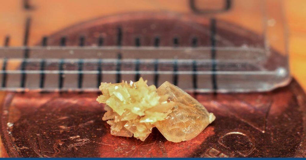
Magnesium ammonium phosphate, often known as struvite stones, is a common component of urinary or kidney stones (MgNHPO4H2O).
A bacterial upper urinary tract infection (UTI) is the root cause of struvite stones. The bacteria’s (Proteus, Klebsiella, Pseudomonas, Staphylococcus, Xanthomonas, or Mycoplasma) waste product, ammonia, can lessen the acidity of the urine (or make it more alkaline).
More alkaline urine will produce struvite stones. A struvite stone mostly contains calcium carbon-apatite and struvite (magnesium ammonium phosphate).
These stones have a quick growth rate and can get rather big, eventually blocking the kidney, ureter, or urinary bladder. This may occur with minor symptoms initially.
Symptoms
Struvite kidney stones are often associated with urinary tract infections and are larger and more difficult to treat than other kidney stones.
Struvite stones can cause pain and block urine flow, leading to complications such as infection and kidney damage. Treatment includes antibiotics to clear the infection and procedures to remove the stones.
Struvite kidney stones can cause the following signs and symptoms:
- Pain in the side, back, or lower abdomen
- Blood in the urine
- Nausea and vomiting
- Urinary urgency and frequency
- Cloudy or foul-smelling urine
- Fever and chills indicate a urinary tract infection
- Painful urination
If you experience the symptoms mentioned above and suspect that you may have Struvite kidney stones, it is important to seek medical attention. Early diagnosis and treatment can help prevent future complications and improve outcomes.
Diagnosis
Struvite kidney stones can be diagnosed by the following tests:
- Blood Testing: Blood tests may detect the presence of excess uric acid or calcium. The results of a blood test allow your doctor to look for additional medical issues while also monitoring the condition of your kidneys.
- Urine Collection: These tests are run continuously. These can help identify whether the patient is producing an excessive amount of minerals that form stones or too few components that prevent stones from forming. Two urine samples should be taken over the course of two consecutive days, the doctor may advise.
- Imaging: The size and position of a kidney stone can be determined using radiology procedures like X-rays, CT scans, MRIs, and ultrasounds.
- Stone Examination: You might be requested to urinate through a strainer to catch stones you pass. Your kidney stones’ composition will be determined via laboratory analysis. This information is used by your doctor to identify the cause of your kidney stones and their composition and to develop a plan to stop further stone formation.
- Intravenous Urography: An intravenous urography procedure involves inserting a catheter into a vein. This checks the kidneys, ureters, and bladder for any problems using X-rays and a particular dye.
Treatment
Struvite kidney stones must be treated because they can harm your kidney and trigger potentially fatal infections if they become large enough.
Shock wave lithotripsy (SWL) or percutaneous nephrolithotomy are the methods used by doctors to remove these stones (PNL).
Treatment for Small Stones:
- Drinking Water: Drinking up to 2 to 3 quarts (1.8 to 3.6 liters) of liquid daily can dilute your urine and help prevent kidney stone development. Drink plenty of water until your urine is clear unless your doctor instructs you otherwise.
- Pain Relievers: For the passage of small stones, your doctor might prescribe ibuprofen or naproxen sodium to treat minor pain.
- Medical Therapy: You might receive medicine from our doctor to aid in passing your kidney stone. An alpha blocker, a class of drugs, relaxes the ureter’s muscles to aid swift kidney stone passage. Tamsulosin (Flomax) and the medication cocktail dutasteride and tamsulosin (Jalyn) are examples of alpha-blockers.
Treatment for Large Stones:
- Shock Wave Lithotripsy (SWL): The stones are broken up into manageable pieces using powerful shock waves from a machine outside the body. The stone fragments will eventually transit through the urinary tract and end up in the urine. Large or numerous stones can need more than one round of therapy.
- Percutaneous Nephrolithotomy (PNL): When SWL cannot remove a patient’s big stone, PNL is the recommended treatment. Scope and other tiny equipment are introduced after a small incision is made in the back. The stone is then removed through the skin wound. The procedure will be performed with the patient sedated. He might need to stay in the hospital for a few days to recover after the procedure.
- Parathyroid Gland Surgery: The parathyroid glands can become hyperactive and lead to the formation of calcium phosphate stones. Your calcium levels may become excessively high due to hyperparathyroidism when these glands create too much parathyroid hormone, which may lead to kidney stones. When a small, benign tumor develops in one of your parathyroid glands, or you get another illness that causes these glands to release extra parathyroid hormone, you may experience hyperparathyroidism. Kidney stone growth is stopped by removing the growth from the gland. Alternatively, your physician might advise treating the illness causing your parathyroid gland to create too much hormone.
- Ureteroscopy: Your doctor may insert a ureteroscope into your ureter through your urethra and bladder to remove a tiny stone from your ureter or kidney. Once the stone has been found, specialized instruments can either snare it or smash it into fragments that will pass through your urine. After that, your doctor might insert a tiny tube (a stent) into the ureter to reduce swelling and encourage recovery. During this treatment, general or local anesthetic may be required.
Prevention
Your doctor might suggest specific medications after surgery to treat struvite stones in the future. Acetohydroxamic acid (AHA) is employed to prevent the bacteria from producing ammonia, which can lead to the development of struvite stones.
Following the removal of a stone, you might also be given antibiotics for some time. Future UTIs might result in stones; thus, this may help avoid them.
Drinking enough water daily will help keep your urine less concentrated with waste items, which is vital for maintaining general health.
If you are well hydrated, your urine should be light yellow to clear since darker pee is more concentrated. It could be advised to consume enough liquids to produce at least two liters of urine each day.
If you have advanced kidney illness, fluid limits may be necessary, so talk to a healthcare provider about how much water is best for you.
Conclusion
Consult a doctor if you experience back and side pain, fever, or frequent urination. Your doctor can do tests to determine whether you have urinary stones and what kind you have.
Most struvite stones can be removed using procedures like PNL and SWL, especially if the stones are tiny. After surgery, there can still remain pieces of big stones. Some patients will require additional surgeries or therapies.
For further assistance on the diagnostic tests for struvite kidney stones, please reach out to HG Analytics. You can also book a consultation with us to know more about the advantages of diagnostic tests pre and post-symptoms of struvite stones.
More On Kidney-Related Issues:
- Can Kidney Stones Kill You? Symptoms, Types & Treatment
- Stage 2 Chronic Kidney Disease
- How Long Can You Live With One Kidney?
- Bruised Kidney – Symptoms, Diagnosis, and Treatment
- Chronic Kidney Disease And Its Stages
- Kidney Scarring: Symptoms, Diagnosis, and Treatment
- Is Pomegranate Juice Good for the Kidneys?


